In this week’s blog, find out how conservators from the National Library of Scotland and National Museums Scotland have been using scientific analysis to discover more about a letter by famous explorer David Livingstone…
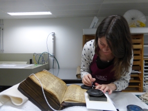
The National Library of Scotland’s collections include a considerable number of letters by David Livingstone, most of which are written a on a white laid paper with an address embossed on the top right corner of the first folio. One of these letters is strikingly different from the others in its appearance, and the reasons for this were recently investigated in a collaborative heritage science project between the Library and National Museums Scotland. The project team comprised Lore Troalen and Jim Tate from NMS and Isobel Griffin and Simona Cenci from the National Library of Scotland.
The letter in question (Acc.13333) (Fig.1), dated 16 April 1865, is written on a very brittle paper that shows a dark, uneven colour. Apart from the colour, this paper appears to be the same as the white paper used by Livingstone for the other letters he wrote around this time, with an embossed address in the same place. This suggests that the dark colour may be the result of chemical degradation, rather than the way the paper appeared originally. However, the discoloration is more severe than the yellowing which often occurs as paper ages, for example as a consequence of exposure to light.

Acc.13333 showing the brown colour of the paper
Scientific analysis was proposed to investigate the nature of the degradation processes that lead to the brown discolouration, which would be of general interest and of practical use in determining whether or not to attempt to remove the discolouration. Additionally, it was hoped that some information about the various coloured inks in the letter might be obtained. The analysis was undertaken non-invasively using X-ray fluorescence (XRF) and Scanning electron microscopy in Backscattered mode/energy dispersive X-ray Spectroscopy (SEM-BSC/EDS), with the SEM in Envac mode.
Results
XRF results
The analysis of the ink with the XRF was problematic, because the beam size (about 2 mm x 1.5 mm) was larger than the ink marks, making it difficult to separate the ink from the paper surrounding it, not to mention the paper underneath the ink. However, the results suggested that all of the inks seemed to be iron based, with traces of copper and zinc, and possibly more zinc in the brown ink.
With regards to the paper itself, the XRF showed significant amounts of sulphur and iron, with the iron present in higher concentrations in the darker areas of the paper. This was interesting because in the previous analysis of various other papers of a similar age, NMS has not detected iron at comparable levels.
SEM/EDS results
The SEM-EDS analysis confirmed the presence of iron in the ink that was tested, as shown in the images below.

The area of ink which was analysed using SEM-EDS


Image above: A detail of an area of writing; the green colouring shows the presence of iron, corresponding to the places where ink is present, and showing that the greatest concentration is where two strokes of the pen overlap
Sulphur was also present in the ink, as seen in the spectrum below. Other elements present in smaller amounts were calcium, sodium, magnesium, aluminium, silicon, phosphorous and potassium, also with zinc in some samples. All samples showed high levels of carbon and oxygen.
With regards to the paper, the elements detected were iron, sulphur, magnesium, aluminium, silicon and calcium, again with carbon and oxygen.

EDS spectra for an area of ink (red) and an area of paper (yellow). The elements zinc (Zn), potassium (K), sulphur (S) and iron (Fe) are notably higher in the ink.
The SEM images of the surface of the paper at high magnification showed in fascinating detail how the ink cracks as it dries out, forming a surface layer which partially obscures the paper fibres.
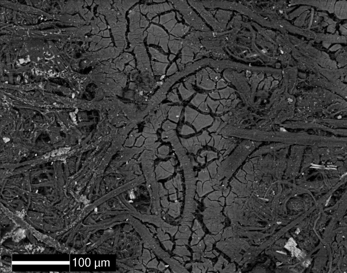
SEM-BSC micrograph of a rectangle of the paper measuring about 0.6mm across, showing a line of ink running up the centre
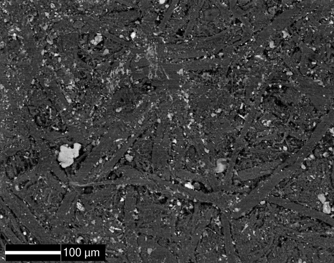
The SEM images also show small and generally angular particles present amongst the paper fibres. The fact that these particles show up brightly tells us that they contain elements which are different from the paper fibres.
Discussion and conclusions
Calcium, iron and sulphur are known to be commonly found in iron gall inks, with the concentrations of other elements varying depending on the provenance of the ink[1]. Given that calcium, iron and sulphur as well as zinc were all found in significant quantities in the inks tested, and that the inks are brown in colour, they seem very likely to be iron gall inks.
It is possible that some of the elements found in the paper may be from water soluble components within the inks, dispersed through the paper by wetting, although this seems unlikely to be causing such a general brown colour throughout the paper.
Various reasons for the dark brown colour of the paper were proposed by the project team:
- It could be due to darkening with smoke, which seems plausible because sulphur as well as carbon is present;
- Since iron is present, it could be due to particles of iron oxide, which were possibly introduced through storage of the letter in a damp and rusty container;
- It could be an organic coating such as oil or wax, which would be difficult to detect with XRF or SEM/EDS; such coatings were sometimes applied to make paper more translucent, although a translucent paper would generally have been used for a copy of a letter and this letter seems unlikely to be a copy because it has an embossed address.
If the small, angular particles seen in the SEM images are the substance causing the brown discolouration of the paper, their appearance makes them more likely to be a sooty material or iron oxide than an oil or wax. However, it is also possible that they are a calcium compound introduced during the manufacturing of the paper.
Hence the analysis raised as many questions as it answered, and there were many suggestions for further testing, which could include:
- Further SEM-EDS analysis, to identify the elements present in the small, angular particles seen in the SEM images;
- Preparation of replicas to introduce soot and iron oxide, followed by analysis of the replicas and comparison of these results to the results for the original letter;
- SEM imaging of some of the other letters written by Livingstone at around the same time, followed by morphological analysis of the images to establish whether all of the letters were written on the same type of paper[2];
- Analysis of the letter to look for organic oils and waxes, for example with Fourier transform infrared spectroscopy. Although it seems unlikely that the paper was coated with oil or wax, it would be helpful to eliminate this as a possibility.
If resources permit, we will tackle some of this analysis in the future, and the mystery of the brown discolouration may be solved. Until then, the letter will remain untreated. And who knows – perhaps it will remain that way forever as a testimony to the letter’s unusual storage history, be that in a smoky room or a rusty old box!
For more interesting conservation conundrums and heritage science research, please follow the National Library of Scotland and National Museums Scotland on Twitter (@NLSColl_Care; @ConserveNMS).
[1] García, J.A., Ruvalcaba Sil, J.L. and Meeren, M.V. (2014) ‘XRF Study of Mexican Iron Gall Inks: Historical and Geographical Overview of their Chemistry’, MRS Proceedings, 1618, pp. 31–41. doi: 10.1557/opl.2014.453.
[2] Kazuyuki, E., Masato, K., Masuchika, K., Barnard, M., Matsuoka, K. and Whitfield, S. (2007) ‘Analysis of morphology and elements on the paper specimens of the Stein collection of the British Library’, in Tradition and innovation: proceedings of the 6th IDP conservation conference, eds. L. Shitian and A. Morrison, National Library of China, Beijing, China, pp. 37-51.